
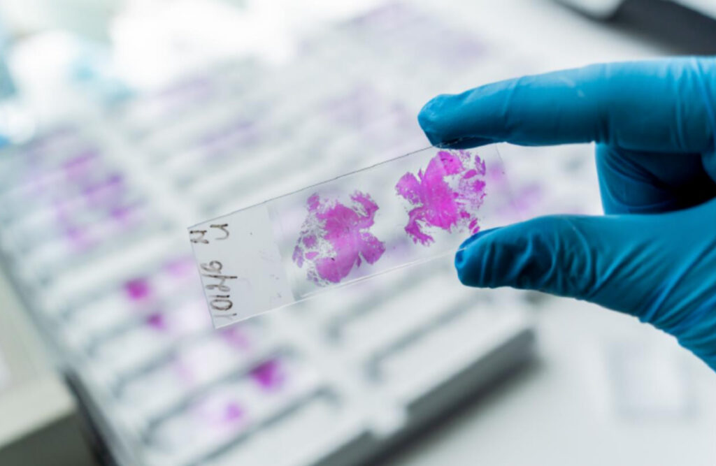
In clinical research and medical product development, biomarkers, imaging, adverse events, and statistical outputs often drive decision-making. These are all essential—but they don’t always show the full picture. They rely on indirect measures, such as lab values, imaging, and performance outcomes, to interpret what might be happening biologically at the treatment site.
That’s where histology comes in—and where many teams either gain real insight or miss the opportunity entirely.
Histology is the study of tissues at the microscopic level. By preparing, staining, and examining thin tissue sections, scientists and pathologists can directly evaluate cellular structure, tissue architecture, inflammation, injury, healing, disease progression, and treatment effects.
In simple terms, histology lets you see what’s actually going on inside the body—not just what other test results suggest.
For teams working in preclinical research, medical devices, pharmaceuticals, biologics, or combination products, histology isn’t just supporting data—it’s one of the most powerful ways to understand safety, mechanism of action, and tissue-level response.
What Histology Evaluates
At its core, histology answers a simple question: What is the tissue actually doing in response to an intervention?
It shows whether tissue is behaving as expected—or if something else is happening, like damage, inflammation, adaptation, or no response at all.
Depending on the study goal, histology can reveal:
- Tissue injury or irritation
- Inflammation and immune activity
- Fibrosis or scarring
- Cell death (necrosis)
- Changes in blood vessels
- How tissue integrates around an implant or device
- How healing progresses over time
- Tumor behavior and changes in the surrounding environment
- Treatment effects—both intended and unintended
- Local tolerability and biocompatibility
A histology slide isn’t just an image—it’s direct evidence of what’s happening biologically. The real limitation isn’t the data itself, but whether studies are designed to fully use it.
How the Histology Process Works
The process itself is well established—but how well it’s executed is what really makes the difference.
1. Tissue Collection
Samples need to be collected from the right location, at the right timepoints, with a clear purpose. If collection isn’t thoughtfully planned, the data may not answer the questions the study set out to address.
2. Preservation (Fixation)
Fixation is a critical control point. Delays or inconsistencies can permanently compromise the sample.
3. Processing and Sectioning
Embedding and sectioning must preserve tissue integrity. Artifacts introduced at this stage can lead to misleading interpretations.
4. Staining
H&E (hematoxylin and eosin), the most commonly used stain to visualize overall tissue structure, is the starting point—but many teams stop there when more insight is needed. Targeted stains and immunohistochemistry (IHC) can reveal much more specific biological detail when aligned with study objectives.
5. Slide Evaluation
Interpretation is what ultimately makes the data meaningful. Whether results are qualitative, semi-quantitative, or scored, their value depends on how they’re evaluated—and how those findings are used.
That’s why histology is not just a lab step—it is a critical part of how study results are understood, interpreted, and acted on.
Why Histology Matters in Product Development
Histology is powerful because it reduces guesswork, it connects hypotheses to biological reality.
Yet in many studies, it’s still treated as confirmatory—or even optional. That’s a missed opportunity.
- Pharmaceuticals: Histology shows whether a therapy is actually modifying disease biology—not just shifting biomarkers.
- Medical devices: It reveals how tissue is responding—whether it’s integrating, tolerating, or reacting—insight that performance data alone can’t provide.
- Biologics and combination products: It highlights immune responses and local effects that often determine long-term success or failure.
In preclinical development, histology should be a decision-driving dataset—not a retrospective add-on.
It can—and should—inform:
- Dose selection
- Device and material design
- Risk characterization
- Mechanism of action
- Go/no-go decisions
Without it, teams are often making high-stakes decisions without a complete view of what’s happening biologically.
What Research Teams Should Ask Earlier
To get the most value from histology, teams need to think about it early in study design:
- What decision do we need histology to support?
- What tissue response would change our development path?
- Are collection timepoints aligned with the biology we expect to observe?
- Do we need H&E alone, or would special stains or IHC provide deeper insight?
- How will these findings connect with imaging, biomarkers, functional outcomes, or safety data?
- Who needs to be involved early to ensure the approach is fit for purpose?
These questions help shift histology from a routine endpoint to a strategic tool.
Final Thought
Histology starts with a tissue sample—but its impact goes far beyond that.
It’s one of the few ways to directly see what’s happening inside the body. And when so many decisions rely on indirect data, that kind of clarity is essential.
The teams that get the most value from histology don’t just include it—they build around it. They ask better questions, choose meaningful time points, and use what they learn to guide decisions along the way.
If the goal is to truly understand how a product interacts with the body—not just whether it appears to work—histology shouldn’t play a supporting role, it should be central.